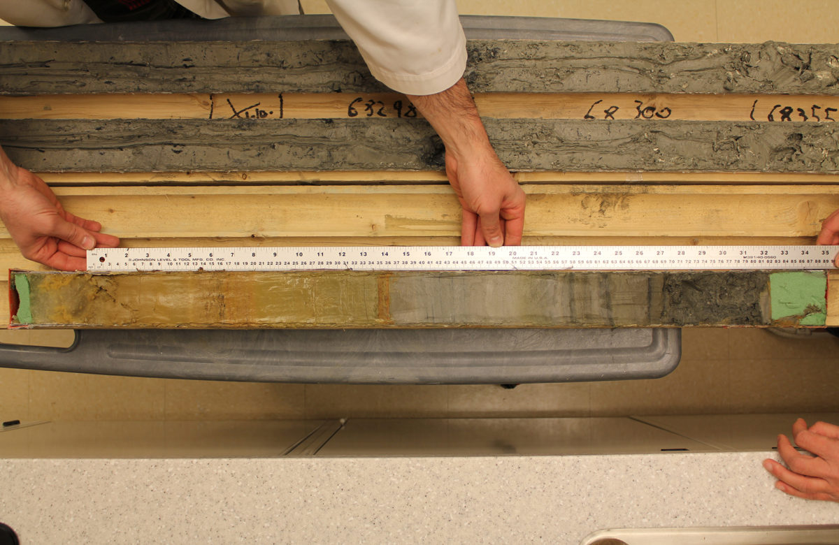
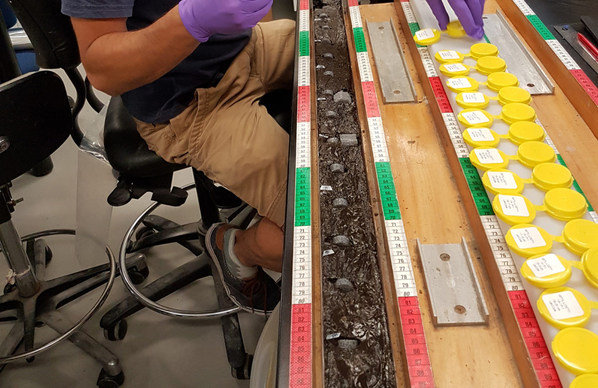
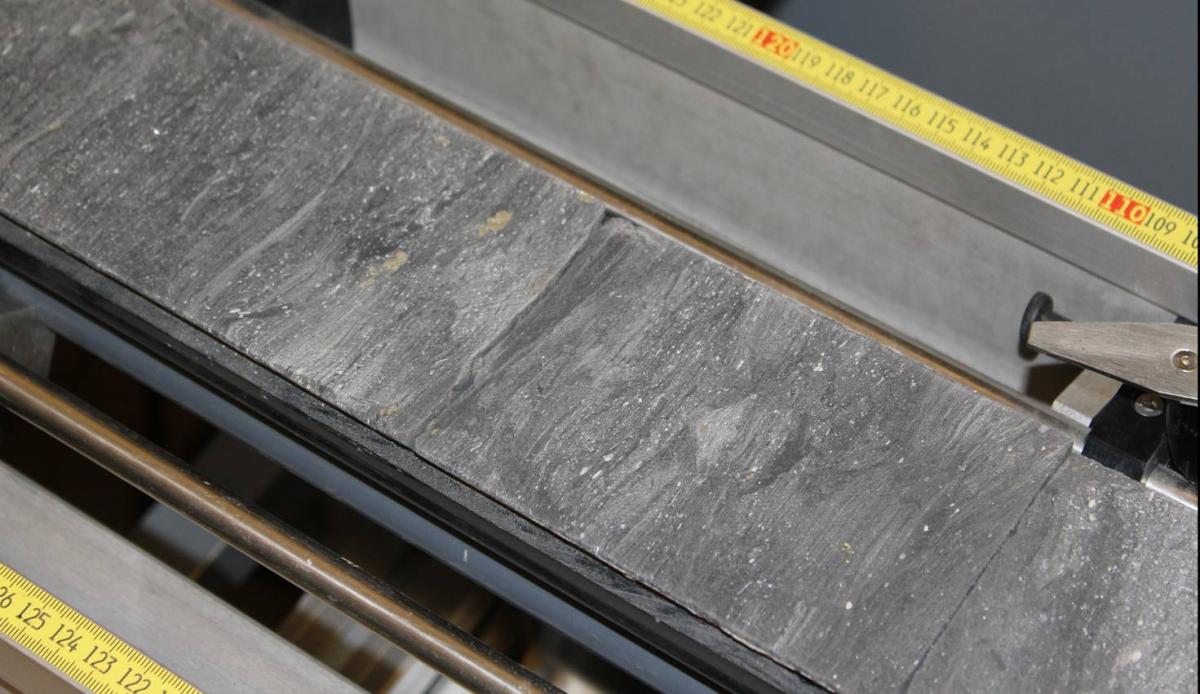
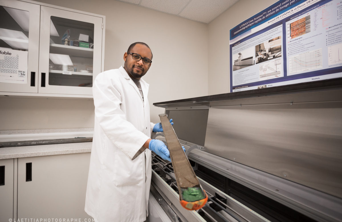
The Geochemistry, Imaging, and Radiography of Sediments Laboratory (GIRAS) generates high-resolution geochemical and radiographic data from marine and lake sediments using non-destructive analysis methods.
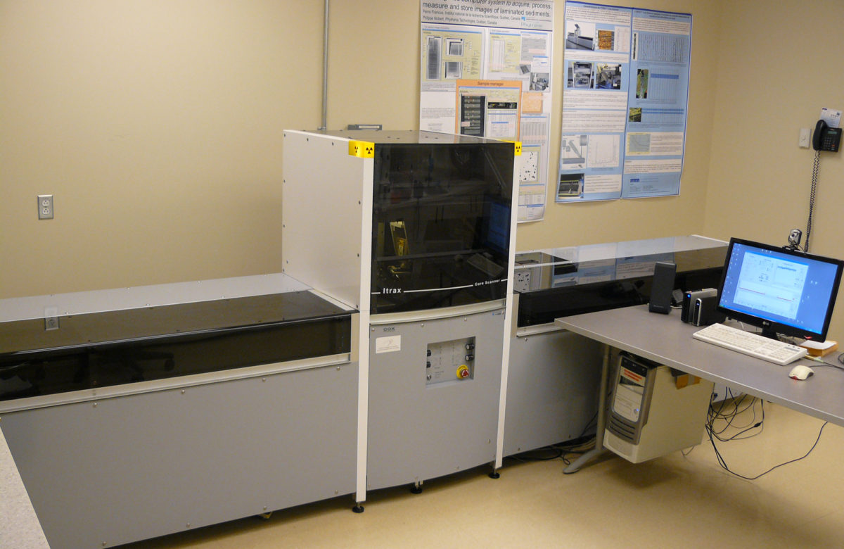
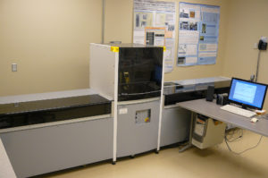
The main piece of equipment is the ITRAX Core Scanner, a non-destructive tool using ultra high-resolution (100 µm) micro X-ray fluorescence for radiographic and chemical analysis of rocks and sediments.
It performs continuous and relatively rapid micro X-ray fluorescence analysis and microradiography of the sample (half-core sections, U-channels, etc.) and records and displays profiles of geochemical changes in sediments.
ITRAX Core Scanner
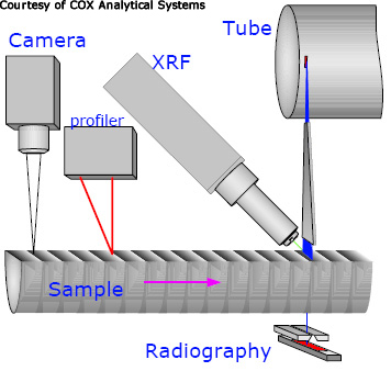
The ITRAX Core Scanner was developed by Cox Analytical Systems. It simultaneously detects microvariations in the density (microradiography) and chemical composition (X-ray microfluorescence) of the sample using two separate X-ray detection systems.
An integrated optical line scan camera provides images of the sample. The analysis is performed without contact with the sample surface and is completely non-destructive.
 |
 |
X-ray source and detection systems
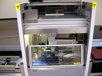
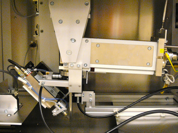
An intense X-ray source (3 kW at 50 mA) is provided by an X-ray tube with a molybdenum anode. The X rays are channeled through an optical device developed by Cox Analytical Systems. The system generates a rectangular beam of a nominal size of 22 mm x 100 microns. Two other tubes are available to improve detection limits for elements from Al to Ti (Cr anode) and for Mo and Nb (Rh anode).
 |
 |
X-ray microfluorescence analysis
Chemical profiles along the sediment core can be recorded for a wide range of elements. The concentrations of these elements can be determined simultaneously by X-ray fluorescence (from Al to U for the molybdenum tube) for concentrations of up to 20 ppm for most elements, depending on the element studied, the analysis time, and the matrix composition. The X-ray fluorescence radiation emitted by the sample is measured at a 45° angle to the incident X-rays by a Si X-ray detector. The detector moves in relation to the sample in order to follow the topography of the sample surface at a constant distance. Digital signal processing provides an analysis of the chemical elements by energy dispersive X-ray spectrometry. Geometric resolution is excellent thanks to the powerful X-ray flux and a well defined measuring point. The analytical resolution is 100 microns per measuring step, the effective measuring point size for XRF analyses being 0.1 x ~ 8 mm.
Detection limits are a function of the atomic number of the chemical element, X-ray exposure time, and the X-ray tube used.
Microradiography
X-rays transmitted through the sample are recorded by a row of 1,024 diodes, each of which is 25 microns wide. An X-ray scan of the sediment core provides undistorted, high-resolution radiographs with details down to 25 microns (pixel size is 0.1 x 0.025 mm; 0.1 mm in the direction of the scan). The radiograph reveals density variations below the percentage and has a very wide dynamic range thanks to the 16-bit images. The successive acquisition of radiographic lines perpendicular to the sample minimizes blurring and distortion commonly found at the corners of conventional radiographic images, providing very high quality images. The system records information line by line as the sample moves through the X-rays. Image quality and resolution can be controlled by selecting the appropriate voltage and current intensity, measurement step size, and exposure time (up to 1 s).
Optical scanning camera
The new high-resolution optical scanning camera system consists of an RGB colour camera operating in successive lines. The camera is synchronized with the motor controlling the step-by-step movement of the sample. The light source uses polarizing filters to mask reflections from the water on the sediment surface. The spatial resolution of the image is 50 µm/pixel.
Magnetic susceptibility probe
The magnetic susceptibility probe in the ITRAX is fully automatic. It moves down to take the measurement and then moves up again as the sample advances. The effective detection area is a 3.8 x 10.5 mm rectangle, which allows high-resolution analysis of the samples.
Sample characteristics
 |
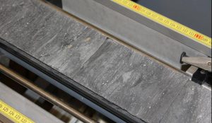 |
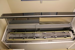
The maximum measurable length of a sediment core is 1.8 m. Sample thickness for XRF analyses can vary from 20 to 80 mm. Maximum sample thickness for a density analysis is 50 mm. These values apply to the sample and not the full diameter of the core. Half-cores, U-channels, rock slices, drill cuttings, and powder samples can be analyzed. For more information, please refer to the section on th ITRAX Core Scanner.
GIRAS facilities and staff expertise are available for external collaborations and research and development contracts. Please contact us for more information.
2025 Rates
| Machine hourly rate (/h) | Handling (/batch)* | Reevaluation (/core)** | Additional assistance(/h)*** | |
| INRS | 30,70 $ | 69,20 $ | 86,50 $ | 69,20 $ |
| Collaborator | 38,20 $ | 86,50 $ | 108,10 $ | 86,50$ |
| Academic | 45,80$ | 103,80 $ | 129,70$ | 103,80 $ |
| External | 61,10 $ | 138,40 $ | 173,00 $ | 138,40 $ |
* These costs include sample preparation, adjustment of acquisition parameters, and delivery of raw data (photos, radiographs, and raw chemical analysis data, expressed in terms of energy dispersal surface). These rates correspond to approximately one hour of work.
** These rates include the re-evaluation of energy dispersion spectra and transformation of raw analytical data from peak area analyses. This step can be done later. It corresponds to approximately 75 minutes of work.
*** For special samples (other than cores), preparation time can be longer. The additional time will be billed at an hourly rate.
Please refer to our Excel table for an estimate of analysis time and cost.
Estimated analysis time for micro X-rays
Each point of analysis has a surface of 100 microns per 8 mm wide (parallel to stratigraphy). If a 1 meter-long core is continuously analysed, 10000 analysis points are obtained. The quality of the analysis increases with x-ray exposure time, particularly for the trace elements. Minimum time is 1 second/point.
| 1 sec per point | 10000 sec/m | 2.8 h/meter |
| 2 sec per point | 20000 sec/m | 5.5 h/meter |
| 3 sec per point | 30000 sec/m | 8.3 h/meter |
| 4 sec per point | 40000 sec/m | 11.1 h/meter |
| 5 sec per point | 50000 sec/m | 13.9 h/meter |
To decrease the duration and cost of analyses, we can make analyses every 200 µm or every 300 µm, etc… It is also possible to combine high spatial resolution analyses but with a shorter acquisition time and a set of more spaced analyses with longer acquisition times for improved analysis quality.
Traditionally, the acquisition of solid-phase geochemical data was time-consuming and required the use of sediments from the sample. The ITRAX makes it possible to acquire high-resolution geochemical data and radiographs from marine or lake sediments without loss or destruction of the analyzed material. Below are some examples:
Marine sedimentsCore from , British Columbia, CanadaThese results include a micro-radiograph, an optical image, and an X-ray microfluorescence (Fe) analysis of a sediment core from Saanich Inlet, British Columbia. This study is part of the IMAGES VIII (International Marine Past Global Changes Study) project conducted on the western margin of the North American continent. Saanich Inlet is a fjord that is 250 m deep and whose entrance is blocked by a sill. It is southwest of Vancouver Island. Restricted deepwater renewal due to the sill, combined with highly productive surface waters promotes conservation of sediments with seasonal varves. In the ITRAX data, volcanic ash from the eruption of Mount Mazama (well-known tephra from 7,645 years ago) is clearly visible at a depth of 139 cm in the core (actual depth: 39.90 m). According to studies carried out under the Ocean Drilling Program (ODP, leg 169S), these sediments were laid down over the last 11,000 years and are made up of alternating homogeneous zones and laminated areas composed of grey to olive-grey diatom mud. (Courtesy of Kinuyo Kanamaru, post-doctoral fellow at the University of Massachusetts, Amherst, Department of Geosciences) |
 |
Lake sediments
Core from Sawtooth Lake, Canada
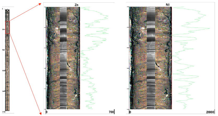
Sawtooth Lake (79°20N, 83°51W) is an oligotrophic lake containing sediments composed of annual varves. The varves are mainly formed during the ice melt season. The average size of the varves is 1 mm, but it can vary from 500 µm et 3 cm. To date, 2,500 years have been measured, which has made it possible to reconstruct past climate variations with annual resolution (Francus et al., 2002). This example of data generated by the ITRAX Core Scanner shows a micro-radiograph, an optical image, and variations in zinc and nickel over a small section of the sedimentary sequence and highlights the resolution capabilities of this instrument.
Francus P., Bradley R., Braun C., Abbott M. & Keimig F., 2002.. Geophysical Research Letters, 29, 20, 1998, doi:10.1029/2002GL015082
Core sample from Lake Yoa, Ounianga Kebir, Chad
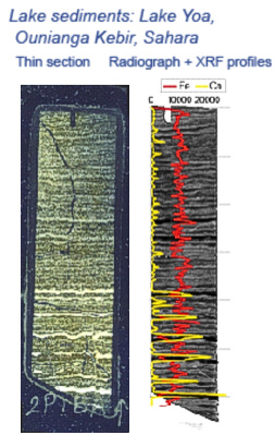
| Lake Yao (4.3 km2; 26 m deep) is a hypersaline lake in northeastern Chad (19.03°N; 20.31°E). It contains thin annual varves (+/- 1 mm) composed of alternating sandy, wind-generated layers, layers rich in organic matter, and layers composed of neogenic micritic calcite.
Thermal stratification of the lake takes place in the summer and is followed by a mixing period during the winter, due in particular to significant variations in seasonal temperatures (Tmax 26-42°C; Tmin 13-26°C) and windy conditions from December to March. This example of data generated by the ITRAX Core Scanner from a small section of the sedimentary sequence shows a combination of an optical image, a micro-radiograph, and iron and calcium geochemical variation profiles. Thin varves show up clearly on the variation profiles, enabling them to be counted. (P. Francus and J.F. Crémer, INRS Eau Terre Environnement Research Centre) |
 |
Rock sample
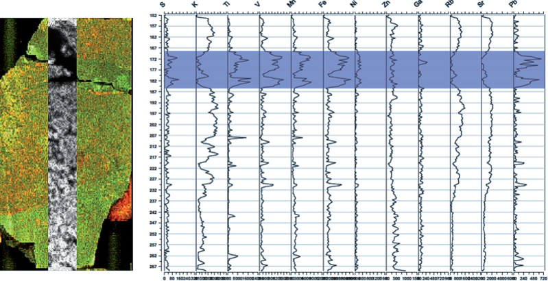
This example from a rock sample analysis shows an optical image, a radiograph, and profiles of chemical variations across areas of hydrothermal alteration. A mineralization zone rich in Ti, V, Mn, Fe, and Pb can be seen. This area is denser than the rest of the sample and shows up clearly on the radiograph (darker area).
Courtesy of Louise Corriveau, Geological Survey of Canada)
This facility is funded by the Canada Foundation for Innovation (CFI), the Government of Quebec, and the Natural Sciences and Engineering Research Council of Canada (NSERC). The Centre de recherche sur la dynamique du système Terre (GEOTOP) is a partner.
Micro-XRF Studies of Sediment Cores
The collective work Micro-XRF studies of sediment cores, edited by I. W. Croudace and R. G. Rothwell, is published by Springer in the Developments in Paleoenvironmental Research collection.
It provides an overview of more than a decade of XRF scanner use and development. These scanners have revolutionized the extraction of high-resolution paleoenvironmental data from sedimentary archives. Professor Francus is highly involved in this scientific venture through his many publications and the acquisition of the initial version of the ITRAX in 2005 (the first in Canada and 3rd in the world), and the latest version in 2012.
Contacts
Arnaud De Coninck
Technical Manager
Phone: 418-654-1801
arnaud.de_coninck@ete.inrs.ca
Pierre Francus
Professor and Scientific head
Phone: 418-654-1801
pierre.francus@ete.inrs.ca
Geochemistry, Imaging, and Radiography of Sediments Laboratory (GIRAS)
Institut national de la recherche scientifique
Eau Terre Environnement Research Centre
490 rue de la Couronne
Québec City, Quebec G1K 9A9
Canada

